Over the next five years, the University of Cincinnati’s College of Medicine is creating a cool cutting-edge space to study the “images of life.”
The way the Center for Advanced Structural Biology (CASB) will do that is through cryo-EM, or electric microscopy. This structural biology methodology gained a lot of attention following the 2017 Nobel Prize awards for chemistry. Cryo-EM is valuable for studying any kind of proteins that are related to any kind of human disease.
What is it and why do scientists do it?
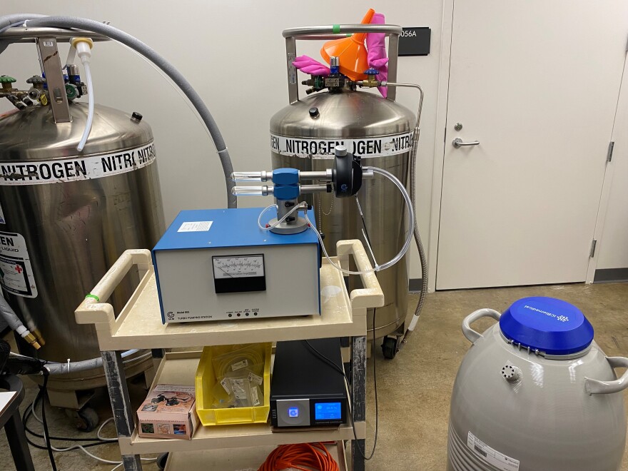
Researchers can prepare and image samples at very cold temperatures to visualize them in a near-native hydrated state. This helps them get a look at proteins at the atomic level.
“We’re actually visualizing a single protein,” says Rhett Kovall, Ph.d., of the Department of Molecular Genetics, Biochemistry and Microbiology, who has helped get the funding and plan the cryo-EM facility in the CASB. “This is quite different from other structural techniques where you don’t get this direct visualization.”
For research scientist and facility manager Desiree Benefield, Ph.d., it’s valuable for studying any kind of proteins that are related to human disease. She first learned about cryo-EM in graduate school.
“I just fell in love with it because you could actually see the science,” she says. “It wasn’t a clear liquid in a tube or a band on a gel — you were looking at your question, which is really exciting to me.”
The samples are flash-frozen with a Vitrobot (specimen preparation unit) and then scientists study them on the Talos L120C Transmission Electron Microscope. In this video, Benefield shows how it all works.
The $1.5 million to buy the equipment came from Research 2030 as part of a JobsOhio grant.
Eventually, Cincinnati Children’s Hospital, the University of Kentucky and Miami University will have access to UC’s equipment.


